Season 2 Case 37
Case 37
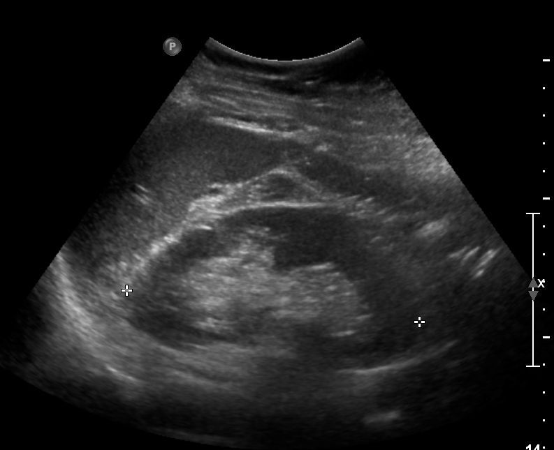
History: right upper quadrant pain
Answer:
CLICK HERE FOR ANSWER
Case 37 Answer:
Looks like I stumped a few of you!
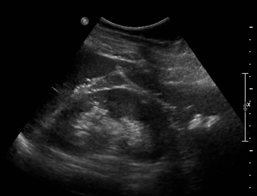
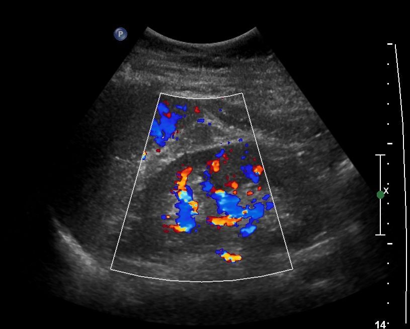
Well the findings, as drawn above (from the same patient) is a mass-like lesion that extended into the renal hilum but does not distort the peripheral renal contour . This lesion is iso-echogenic to renal cortex without increased internal vasculature (the Doppler image shows normal hilar vessels).
Diagnosis: hypertrophied Column of Bertin (also known as “junctional parenchyma” or formerly “septum of Bertin”). - ie normal renal cortex extending into the renal medulla.
here is another example I found online: https://horizon.rad.msu.edu/studyshare/cgi-bin/repos/studyshare_repo/wrm/repo-view.pl?cx_subject=22280479&cx_repo=studyshare_repo
Reference: http://radiopaedia.org/articles/hypertrophied-column-of-bertin


