Season 4 Case 16
Case 16
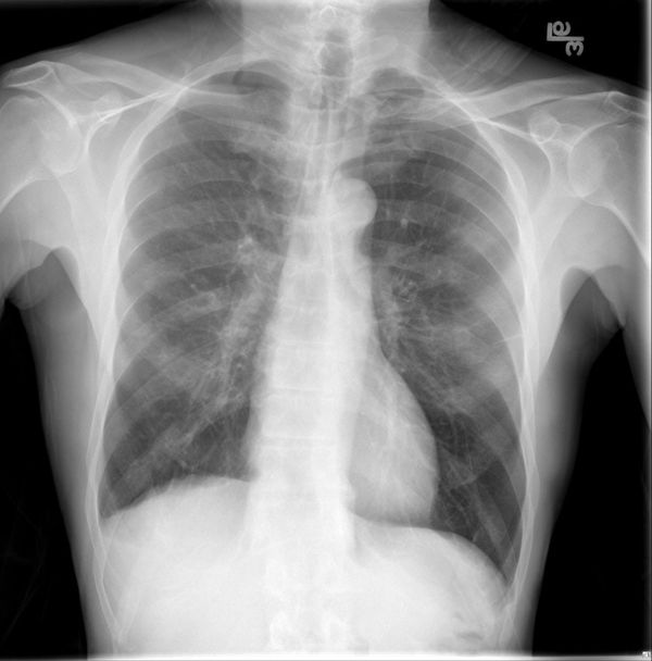
This case is a little different. This patient got X-rays performed at their Internist’s office who diagnosed them with a hilar mass and probable lung cancer. Of course it is Friday afternoon and unfortunately your CT scanner is down for maintenance (isn’t that always the way?). Can you help? What do you think of the Internist’s diagnosis and what Radiology smarts can you use to support your decision?
<
Answer:
CLICK HERE FOR ANSWER
Case 16 Answer
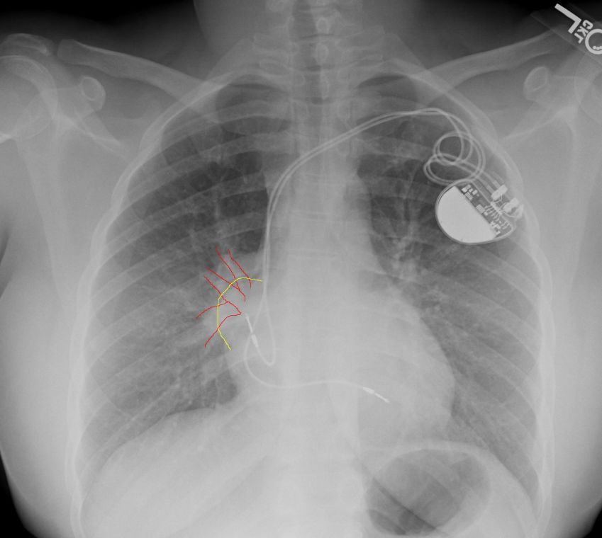
So this may have been a little difficult without the ability to window/level but there are 2 findings here: 1) obscuration of the right heart border and 2) hilum overlay sign (which was the attempted learning point).
Hilum overlay sign is an appearance on frontal chest radiographs of patients with a mass projected at the level of the hilum which is in fact either anterior or posterior to the hilum. So despite seeing a mass “overlying” the hilum you can still see vessels coursing through it, ie you can still see the hilum
http://radiopaedia.org/articles/hilum-overlay-sign
This is in contrast to the hilar convergence sign which shows the hilar vessels converging to the mass but not extending in front or behind it.
http://radiopaedia.org/articles/hilum-convergence-sign
So did we do a CT? you know we did.
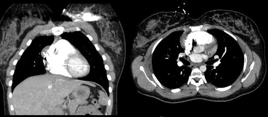
Diagnosis: Pericardial cyst

