Season 8 Case 39
History: altered mental status
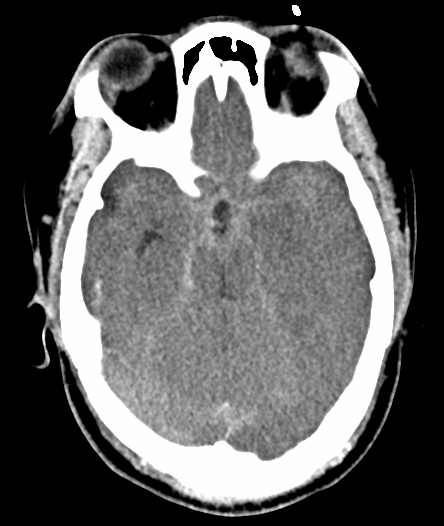
Answer: Psuedosubarachnoid hemorrhage secondary to hypoxicischemic brain injury
In these 2 images we see hyperdensity (increased attenuation) along the basilar cisterns. While this is a common appearance of subarachnoid hemorrhage secondary to Circle of Willis Aneurysm rupture, we need to look at the entire image and the entire exam!
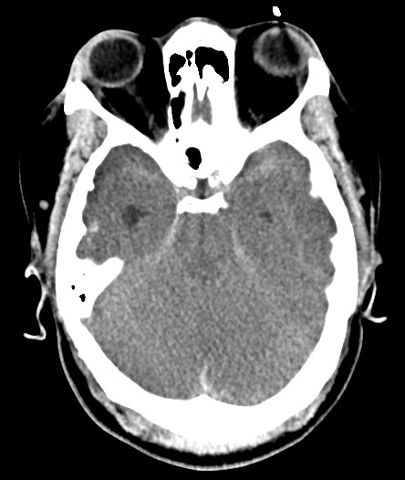
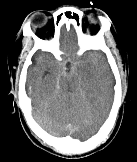
How about the basilar cisterns? - They are effaced
How about the grey-white junction? - It is gone. In fact, Normally grey matter is slightly hyperdense compared to the white matter, but here we have the peripheral grey matter actually lower in density than the central white matter (aka the reversal sign)
How about the central grey matter structures? Similarly, the caudate nuclei, basal ganglia and even thalami are all low in density - which again is not normal
How about the normal sulcal pattern of the cerebrum? - It’s gone. In fact can you even find a sulcus? (and the patient isn’t 15 years old)
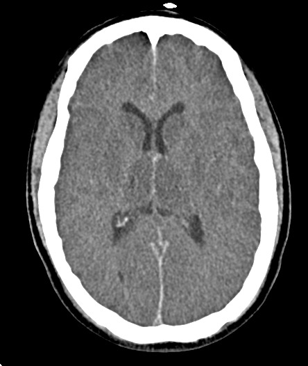
This is an example of global anoxic injury aka hypoxic-ischemic encephalopathy. Global anoxia leads to neuron injury. Since grey matter is more metabolic than white matter, it is effected earlier and more severely. Anoxia/Ischemic -> neuron injury -> edema -> decreasing grey matter density.
So what about that increased density all throughout the basilar cisterns, sylvian fissure, falx and tentorium? While it may look like blood, it’s actually artifact - ie pseudosubarachnoid hemorrhage.
Because of the cerebral edema, particularly the grey matter edema, the parenchymal density decreases. This combined with outflow venous dilation and thrombosis secondary to the increasing intracranial pressure, yields the appearance of increased density particularly throughout the basilar cisterns.
If you can get a good density measurement you will see it is more in the normal 30-40 HU venous density range compared to extra-vascular or true subarachnoid hemorrhage in the 60-70 HU range.


