Season 10 Case 6
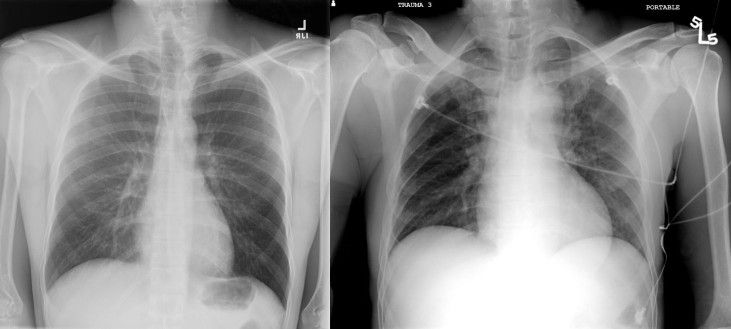
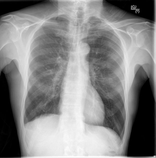
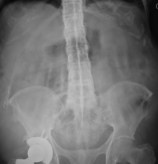
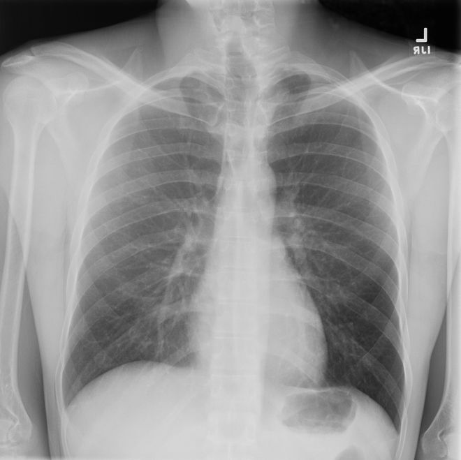
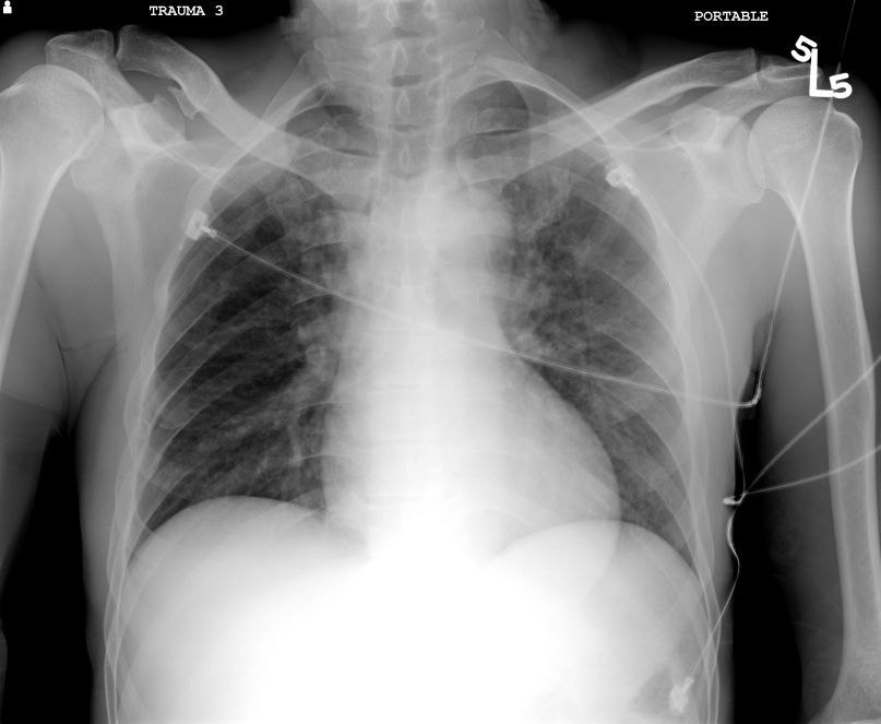
Three Chest X-rays, same finding on all the X-rays below. Can you determine the cause on each?
Answer:
CLICK HERE FOR ANSWER
Answer: Unilateral hyperlucent lung
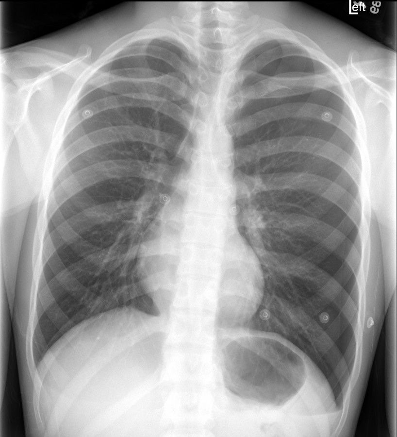
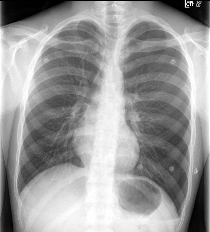
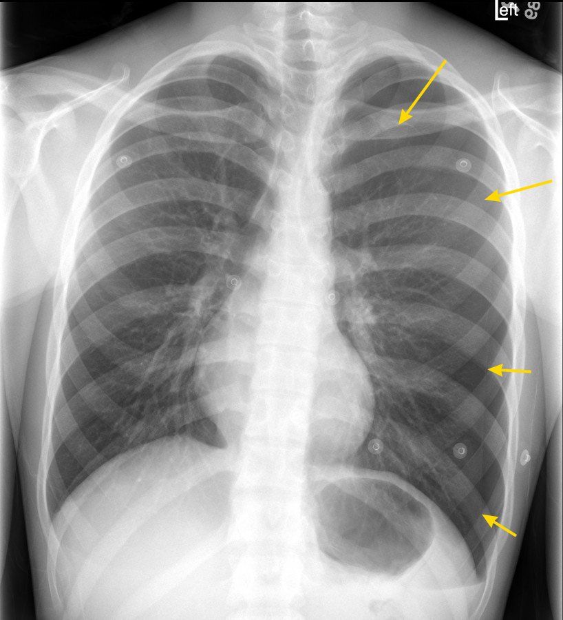
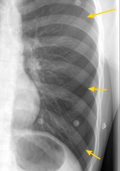
Left: Left pnuemothorax (left side)
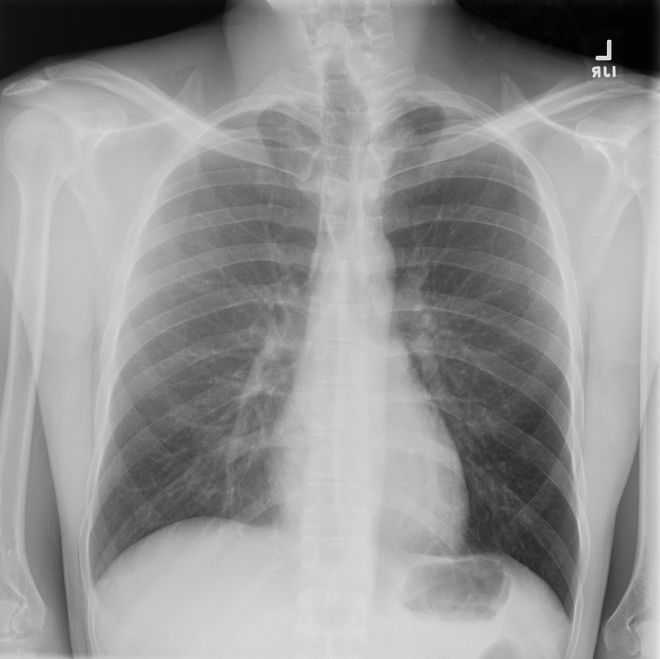
Middle: Grid de-centering arifact underexposing the right>left sides of the chest (left side)
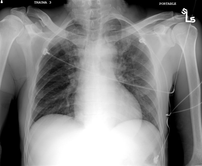
Left: Poland's syndrome with absent right pectoralis muscle (right side)
Left Case:
We have a young healthy patient with an enlarged, hyperlucent left hemithorax. Note the depressed left diaphragm and the mild shift of the mediastinum to the right. I marked the pleural line for you.
?Tension pneumothorax? technically you need cardiovascular compromise for that diagnosis but regardless we are trending in that direction so I would call them! (I prefer not to let it get to cardiovascular collapse but that may be me).
Projection can be difficult with pneumothoraces as that thin white pleural line can be difficult to see, particularly in a young healthy patient. Pneumothoraces tend to be non-dependent and parallel the chest wall as the collapse inward.
Middle Case
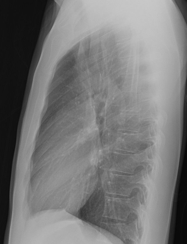
This is just a normal chest X-ray. But due to alignment of the grid collimator we have relatively less photons on the right side of the chest compared to the left. This causes relative underexposure of the right side of the chest yielding a relatively hyperlucent left chest.
Note that everything in the right chest is "whiter", the lung, the bones, the supraclavicular soft tissues and the chest wall. The lateral is normal which helps confirm.
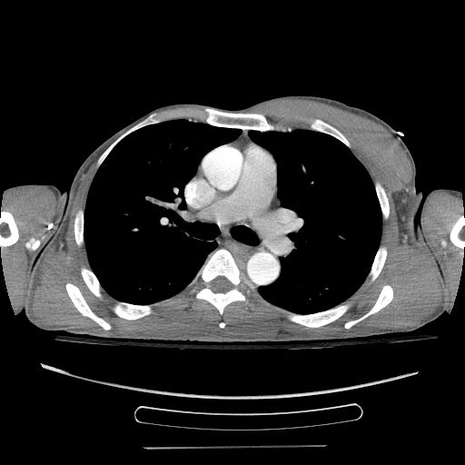
Right Case
This is the fun one. Poland's syndrome is rare congential absence of the pectoralis muscle. Generally sporadic and male = female incidence. It is associated with rib and upper limb abnormalities (so examine the ribs and shoulder girdle). Thus, we have less soft tissue to penetrate with X-rays on that side, leading to a relatively overexposed ispilateral lung. Mastectomy is a much more common cause of the same phenomenon.
The Chext X-ray shows a relatively lucent right lung but the bones and soft tissues look the same at the contralateral side. In fact, even pulmonary vessels are actually better defined on the right due to increased X-ray exposure. CT confirms absence of the right pectoralis muscle.
What reasons can you come up with for differential densities for the hemithoraces?
Just a few:
- Pneumothorax
- Layering pleural fluid
- Artifact
- Positional (rotation)
- Scoliosis
- Soft tissue - Poland's syndrome, mastectomy
- Obstruction ie foreign body (especially in kids)
- Unilateral emphysema

