Season 10 Case 15
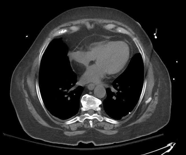
History: Chest Pain
Answer:
CLICK HERE FOR ANSWER
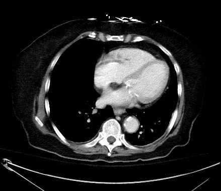
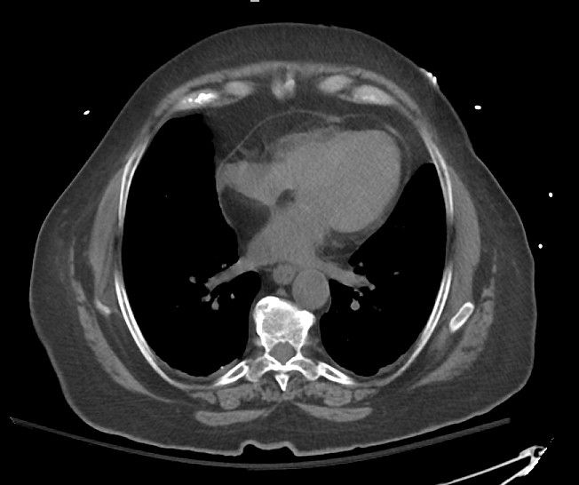
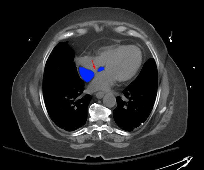
Answer: Lipomatous hypertrophy of the interatrial septum
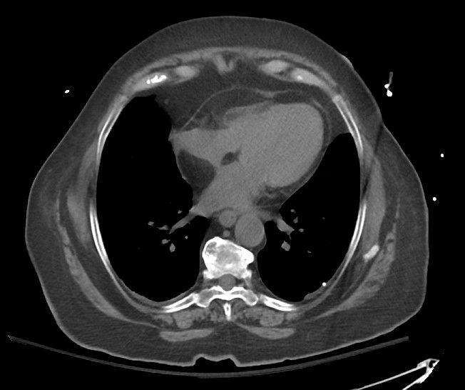
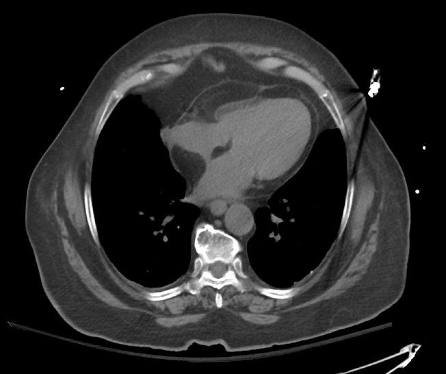
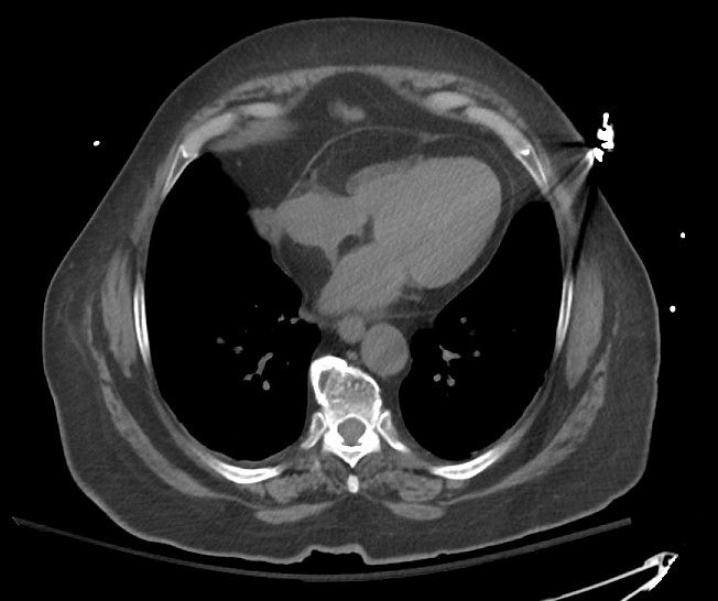
This entity is exactly what the name implies, there is hypertrophy and fat deposition between the fibers of the inter-atrial septum.
- Commonly has a smooth mass-like bulge into the atrium
- usually spares the fossa ovalis (the "closed" depression in the right atrium at the site of a formerly patent foramen ovale during fetal development)
- Does NOT have a capsule (cardiac lipomas do have a capsule)
- associated with mediastinal lipomatosis (excess fat deposition through the mediastinum)
Radiology:
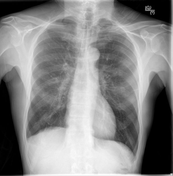
Plain films: not appreciated (perhaps an enlarged mediastinum if there is associated diffuse lipomatosis)
CT: thickened (>2cm), nonenhancing fat (very low) density between the right and left atria. Again, commonly spares fossa ovalis yielding a bilobed or "dumbbell" appearance.
MRI: high intensity on T1, hight= intensity on T2, low intensity on fat-suppressed consitent with fat. Should be homogeneous.
PET: shows + FDG uptake theorized due to brown adipose fat composition (remember, brown fat is FDG avid).
Clinical:
Generally this is a benign and incidental finding and no further work-up or treatement is necessary unless there is significant mass effect on SVC or atrium.
But this has been associated with cardiac dysrhythmias (eg atrial fibrillation, premature atrial contractions and atrioventricular block) as well as syncope and sudden death. So in the setting of cardiac conduction abnormalities, perhaps one may want to suggest Cardiology consultation.
This Patient
In this patient, it was believed their pain was due to an alternate etiology so no further work-up was felt warranted.
The key is to recognize this and differentiate from other cardiac lesions such as:
- cardiac lipoma (usually more capsulated, extraluminal)
- cardiac rhabdomyoma/rhabsomyosarcoma (usually more pedunculated intraluminal lesions, think ventricular, often with calcifications from prior intra-lesion hemorrhage. + enhancement, usually in newborns/very young pts)
- atrial thrombus (purely intra-luminal)
- cardiac myxoma (usually Left atrial, low but not fat density, can have calcification, often move with contraction)
