Season 8 Case 1
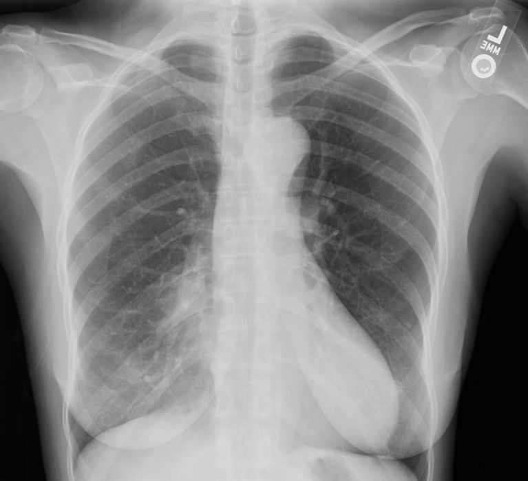
History: Abnormal Chest X-ray?

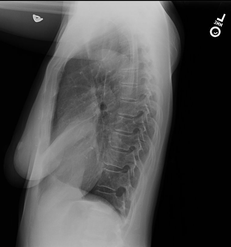
How about a lateral?

Answer: Right middle lobe atelectasis
Note the obscurred right heart border on the frontal. Now this can be difficult as other processes can also so this including pneumonia and even pectus excavatum deformity.
But the lateral shows the narrow triangle overlying the heart, classic for right middle lobe volume loss


