Wetread Case 38
History: chest pain
Diagnosis?

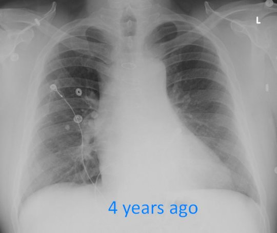
Answer: Penetrating Atheromatous Ulcer (PAU)
What? How you say? Let’s walk through this.
Here’s the current Chest X-ray and I’ve added labels (blue = heart border, red = aorta, orange = hilar vessels).
We see a left infrahilar mass that doesn’t obscure the left hear border (blue) but does obscure the aortic border (red, with dashed lines being where we think it should go and the dark red line outlining the mass). We can still see the hilar vessels (orange) so this mass can not be the vessels nor silhouette the vessels (ie the hilum overlay sign)
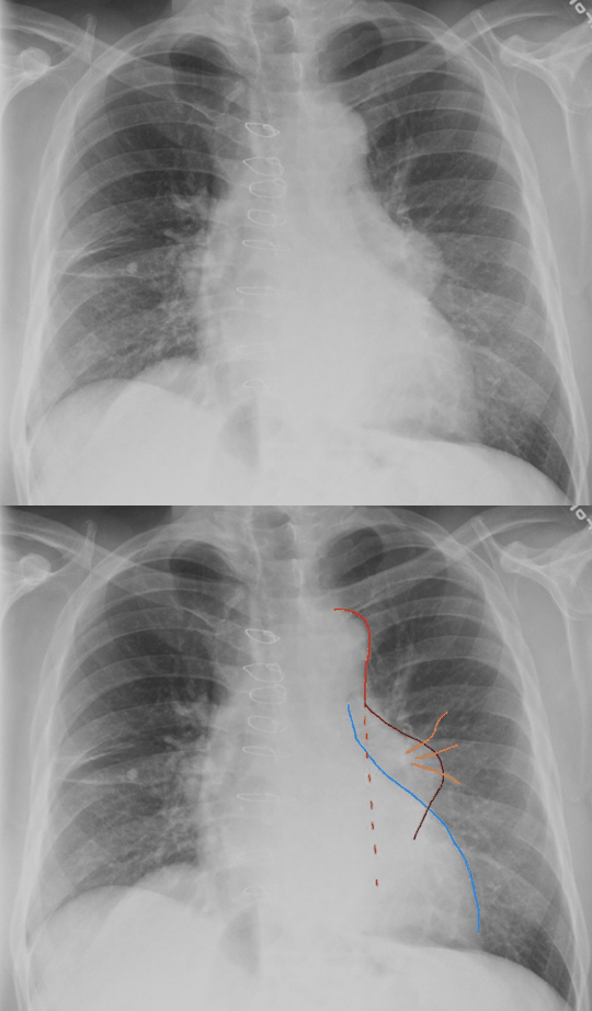
So this mass has to either obscure the border of the descending thoracic aorta or be the descending thoracic aorta. And if it is the aorta, we have a focal mass or enlargement of the aorta.
Can we confirm?
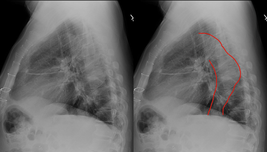
It is difficult to see the entire aorta, but red lines are my guestimate suggesting increased diameter over the mid throacic spine.
So when the answer box (aka CT scanner) comes back up, here is what we see.
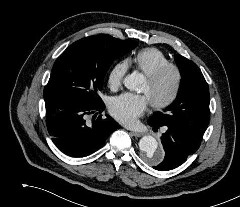
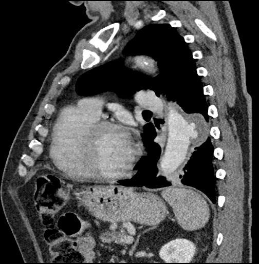
Sure enough there we see focal bulging of the aorta which just so happens to contain contrast in keeping with a PAU.
Aortic wall pathology can be a bit confusing. There are a bunch of good articles including Radiology Assistant from which I am borrowing their wonderful images. I encourage everyone to go read their wonderful write-up. In summary:
Intramural hematoma is isolated blood within the vessel wall with no connection to the inner lumen

Ulcerated plaque is just that, an ulcer in atheromatous plaque but it does NOT extend beyond the intimal layer
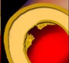
If the ulceration errodes beyond the normal intimal layer into the vessel wall (ie the media layer), now we have a penetrating atheromatous ulcer (PAU)

All of the above can progress to an aortic dissection. So there is injury to the vessel intima allowing blood into the media layer and then the pressure gradient causes extension of the injury through the vessel wall (ie media layer)
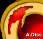
While that may be a bit simplistic, hopefully that helps understand the different types of aortic wall pathology.

