Season 1 Case 35
Case 35
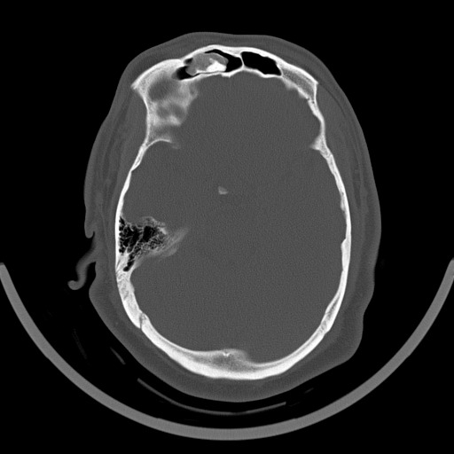
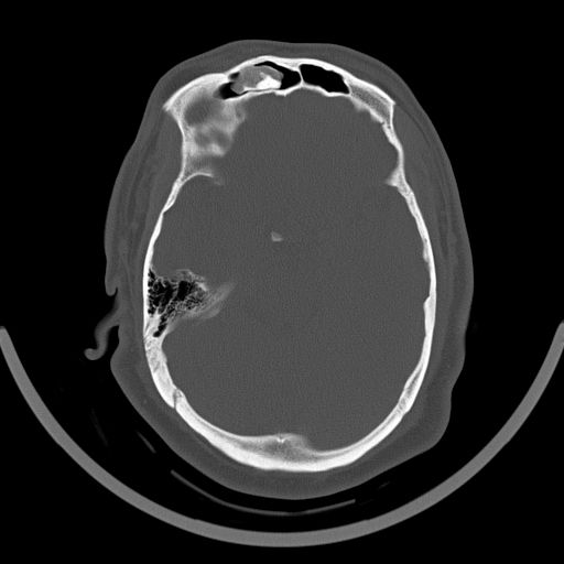
History: head pain/head trauma

Answer:
CLICK HERE FOR ANSWER
Case 35 Answer: Osteoma of the frontal sinus
Seems like an easy one right? Well I think this may be one of those cases you get right if you don’t think TOO much about it. It sounded like there was some concern with the diagnosis of osteoma due to varying density within the lesion. Otherwise it is classic in location and even contains some of the bone-cortex density we classically think of. THUS the reason I put this on here. As it turns out, there are several variations on the classic paranasal sinus osteoma. I have included the short reference below, but just remember that there are actually 3 types (below from the reference):
- ivory osteoma
- most common
- dense bone lacking haversian system
- most common
- mature osteoma
- also known as osteoma spongiosum
- resembles ‘normal’ bone, including trabecular bone often with marrow
- mixed osteoma
- mixture of ivory and mature histology
CT demonstrates a well circumscribed mass of variable density, varying from very dense (similar in density to normal cortical bone) to less dense with a ground-glass appearance. They are seen either with a sinus or less commonly exophytically growing out of a sinus.


