Season 2 Case 18
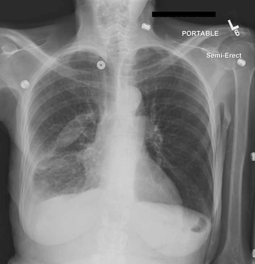
History: chest pain
Answer:
CLICK HERE FOR ANSWER
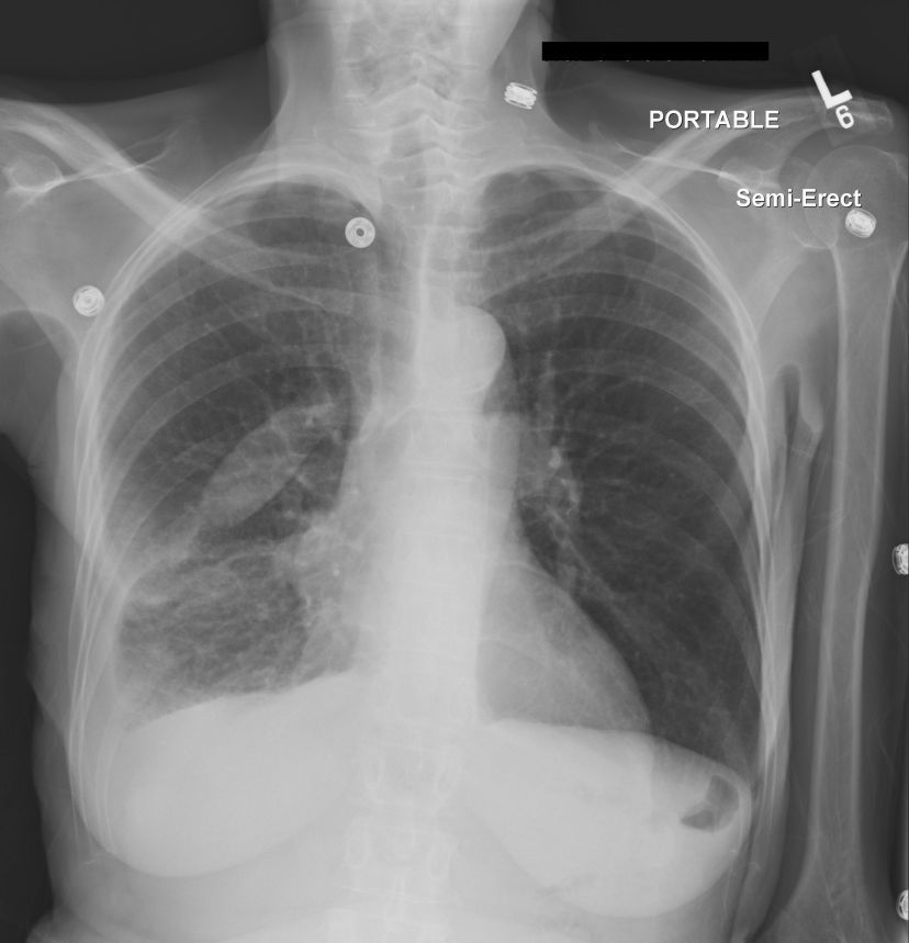
Answer: Pulmonary Pseudotumor
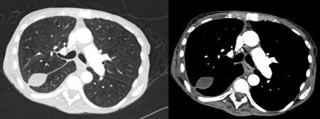
I don't recall as to whether the CT was done to specfically evaluate the "mass" or if it was done for another reason, but it is clear that this "mass" is simply loculated fluid within the right major fissue.
Pseudotumor is exactly that, it's something that looks like a "mass" but is not. There are several examples in the chest:
- loculated pleural fluid
- round atelectasis
- epicardial fat pad
- mucoid impaction of bronchi
The key here is the shape. Classically loculated pleural fluid demonstrates an elongated, oval or biconvex, sausage-shaped, well marginated "mass" along the course of the major or minor fissures (don't forget the accessory fissures - ex azygous vein in the azygous fissue!).
While CT may not be necessary (as in this case), it is not uncommon to progress to CT these days out of lack of experience of out of caution.
The key is to familiarize yourself with these pseudo-masses so one doesn't cause undo concern to the patient/clinicians as well as to save time, energy and expense. But sometimes further work-up is warranted. It wouldn't be the first time I saw CXR mass -> CT peripheral mass (? round atelectasis) -> biopsy showing normal lung tissue.

