Season 2 Case 21
Case 21
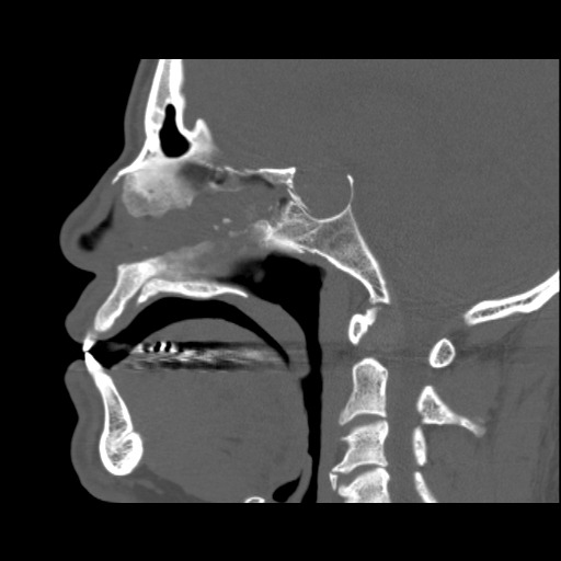
History: trauma
Answer:
CLICK HERE FOR ANSWER
Case 21
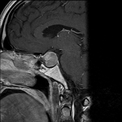
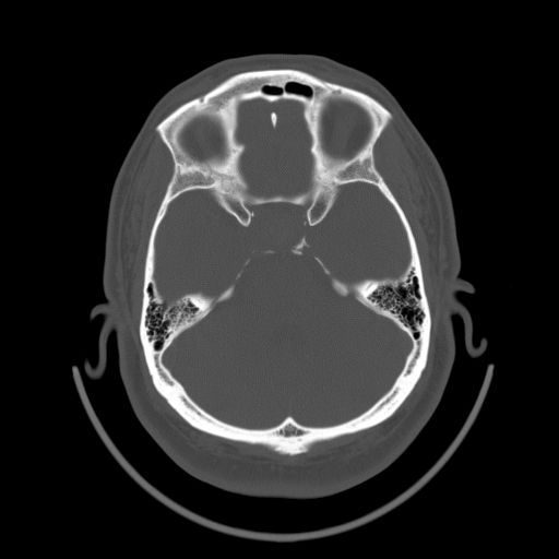
Additional axial CT and sagittal MR images. As many of you noticed, the sella is enlarged. I didn’t really give you enough characteristics to determine fluid or soft tissue density which should sway you from empty-sella to tumor respectively. But you can see on the images I just posted that there is a soft tissue mass there consistent with a pituitary macroadenoma
ref on pituitary macroadnomas: http://radiopaedia.org/articles/pituitary-macroadenoma-1
Upper normal sellar size on radiographs: 17mm AP and 13mm height http://roentgenrayreader.blogspot.com/2011/06/sella-turcica-dimensions-on-radiographs.html
Nice write-up of different sellar/parasellar lesions: http://www.radiologyassistant.nl/en/p485d7745cc720/sella-turcica-and-parasellar-region.html

