Season 2 Case 28
Case 28
History: head injury

Answer:
CLICK HERE FOR ANSWER
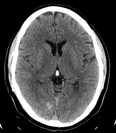
Tricky one with the history (which is why I put it!). The key here is the shape on the CT. It is somewhat amorphous for an interparenchymal hemorrhage, and no noticeable edema around it. The MRI shows a somewhat triangular area of susceptibility artifact (blooming) on the Gradient Echo imaging. All together this is most consistent with a:
Answer: Cavernous malformation
Keys:
hyperdense on CT (50% show Ca++,)
hemosiderin ring (blooming) on MRI (GRE most prominent)
no mass effect
m/c history is seizure or neurologic deficit but they are commonly found incidentally
Sources:
http://www.neuroradiologycases.com/2012/01/cavernoma-cavernous-malformation.html
http://radiopaedia.org/articles/cerebral-cavernous-malformation


