Season 10 Case 1
History: Shortness of Breath
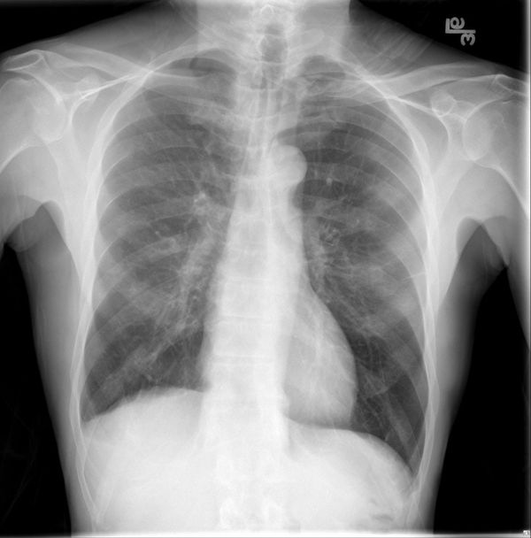
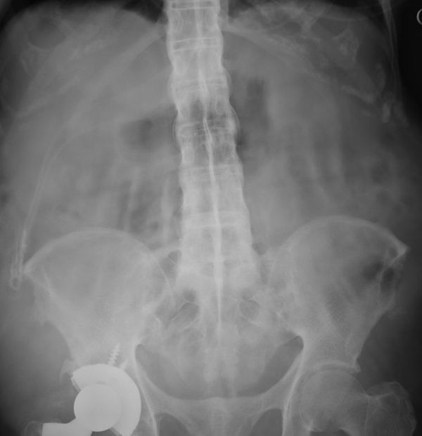
What's the Radiologist's best friend? Prior images of course!
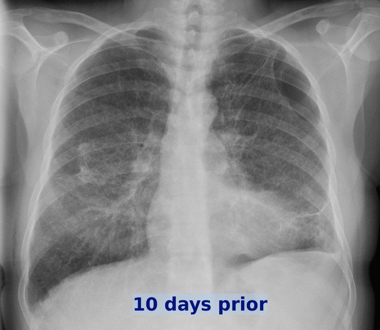
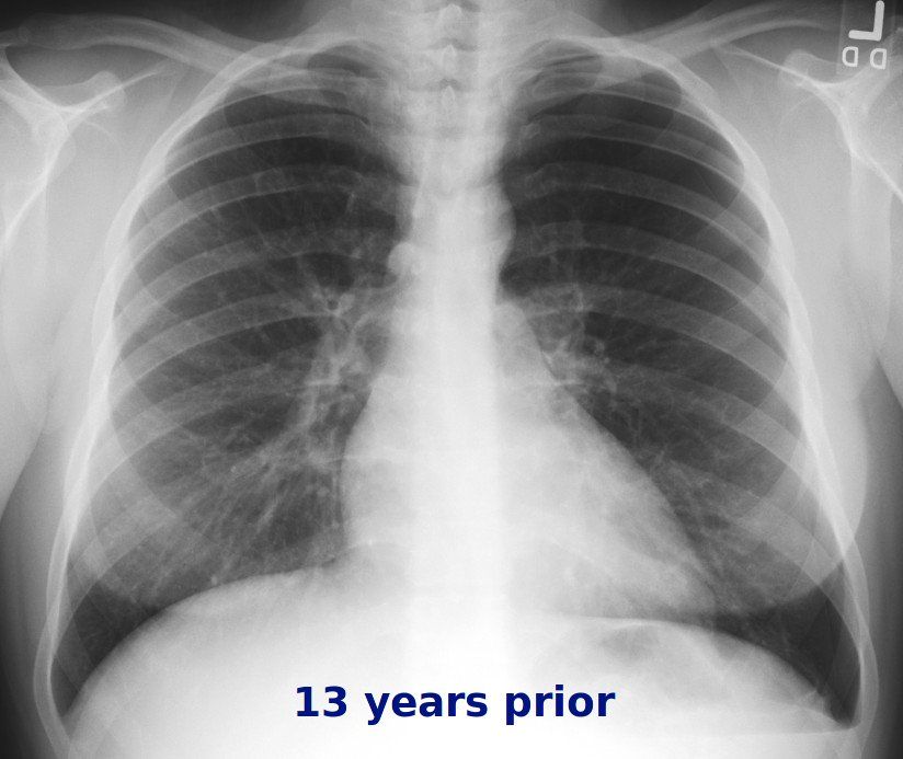
Answer:
CLICK HERE FOR ANSWER
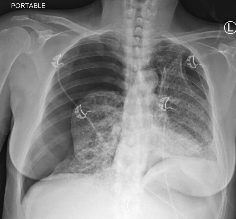
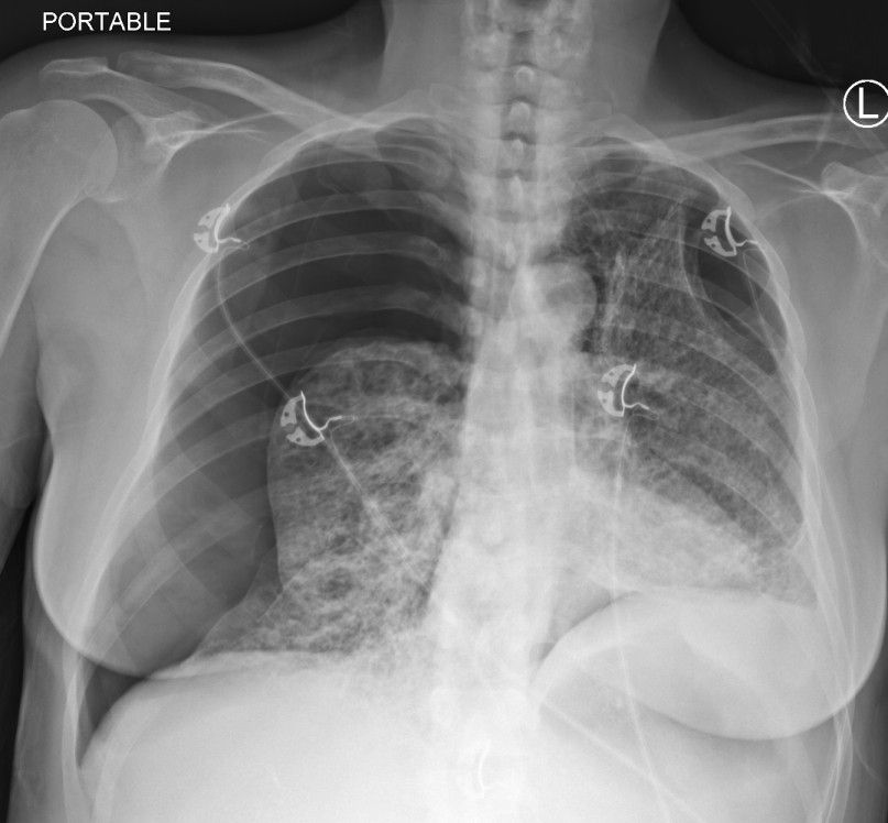
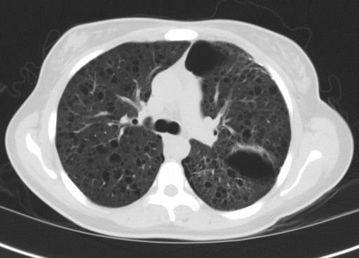
Answer: Pneumothorax secondary to Lymphangioleiomyomatosis (aka LAM)
So we clearly see the large pneumothorax on the right (medially displaced pleural line with absent lung markings peripherally), but how about the cause?
Notice the appearance of the lung, ie the cystic appearance. While there are many causes of cystic lung disease, what do you think the age of the patient is? Yes, it's a middle aged female. Does emphysema make sense? What would make more sense in this patient is LAM.
Cause: LAM is a multisystem disorder characterized by insterstitial proliferation of smooth muscle cells that can obstruct veins, lymphatics and small airways.
Population: Typically in women of childbearing age.
Radiology:
Chest:
- thin-walled cysts of variable size with NORMAL lung parchenchyma
- recurrent pneumothoraces
- chylothorax (blocked lymphatics)
Abdomen:
- Renal angiomyolipomas (#1)
- splenic cysts
- lymphadenopathy
- uterine fibroids (leiomyomas)
- Chylous ascites
So what does the CT show?
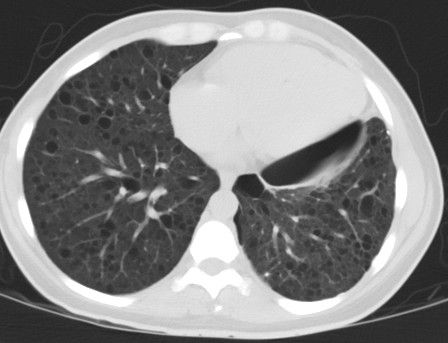
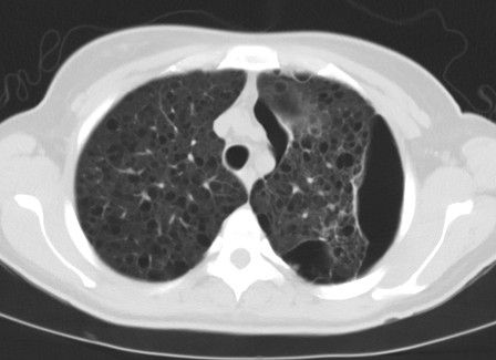
And what do you think about this last image of the CT?
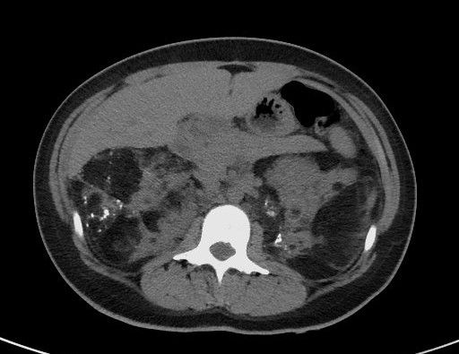
Yep, renal angiomyolipomas (yes, these look much larger and more invasive than the ones we more commonly see spontaneously).

