Season 10 Case 8
History: Neck Pain

Answer:
CLICK HERE FOR ANSWER


Answer: Osteoblastoma (versus osteoid osteoma)
Osteoblastomas and osteoid osteomas are pathologically very similar lesions, radiographically differentiated primarily based on size.
- Osteoid Osteoma: <2cm in size (small lytic component vs sclerosis)
- Osteloblastoma: >2cm in size (larger lytic component)
Both are benign bone tumors
- Osteoid osteomas ~10% of benign bone lesions
- Osteoblastomas ~3% of all primary bone tumors
- classically found in adolescents/young adult (ages 10-35yo)
- osteoblastomas likely skewed to the older ages
- male:female = ~3:1
The lesion itself is a central lytic nidus of osteoblasts, osteoid and vessels. It is surrounded by a fibrovascular rim and surrounding reaction (sclerosis). It is this nidus that releases prostaglandins which is what causes the pain.
Clinical:
- Classic presentation is focal pain in an adolescent, often during the night, that is relieved by aspirin/NSAIDs (* osteoblastomas are typically less responsive to NSAID therapy)
- When in the spine, can cause painful scoliosis that is concave towards the lesion
- If the lesion is at/within a joint, presentation can be confusing for inflammatory arthritis
Location:
- Osteoid osteoma: Long bones > Phalanges > Spine (lumbar > cervical > thoracic)
- Osteoblastomas: Spine (cervical > sacrum), classically posterior elements
Radiology:
- X-ray
- Osteoid Osteoma: Often the small lucent nidus is difficult to see on X-ray, but look for an area of sclerosis/cortical thickening/periosteal rection around the nidus
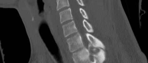
- Osteoblastoma: Larger lytic component more visible, with expansile bubbly appearance, +/- internal calcifications, + surrounding sclerosis
- CT
- Modality of choice
- Osteoid Osteoma: see the sclerosis as well as the central lucent nidus which may have a small central sclerotic "dot"
- Osteoblastoma: similar to X-ray with better characterization of internal matrix +/- calcification. Aneurysmal bone cysts are commonly seen concurrent with these lesions.


Nuclear Medicine
- Back in the day, bone scans were performed and one would see extensive Tc-99m uptake/activity at the lesion ("blastic" lesion)
- Double-density sign - ie area of increased uptake with a central area of even MORE increased uptake corresponding to the nidus
MRI - Signal and enhancement of the central nidus can be variable with large amount of surrounding edema
- Edema sometimes confused for an aggressive lesion
Treatment:
- Osteoid Osteomas can be treated symptomatically with NSAIDS/aspirin (which is somewhat diagnostic for them)
- *remember osteoblastomas tend to be less responsive to antiinflammatory therapy
- Classic treatment is surgical resection
- Newer techniques of radiofrequency ablation are becoming common
- Some believe that small osteoid osteomas can spontaneous resolve over time and thus a conservative course of symptomatic treatement (NSAIDS/aspirin) may be possible (average resolution in ~33mo)
Differential Diagnosis:
- Brodie's abscess (ie osteomyelitis) - central lytic nidus (no ca++ and "cold" on bone scan), but can have surrounding sclerosis (lesser degree). Look for a linear lytic sinus tract extending through the cortex
This Case:
This case falls in the cross-over between the two lesions. It was ~2cm in size, but in an older patient (fused growth plates) and with a more expansile appearance in the posterior elements of the spine. In reality, it probably doesn't matter too much as they both are treated the same except possible response to antiinflammatory therapy.


