Season 10 Case 13
Hx: 27 yo with Shortness of Breath
Answer:
CLICK HERE FOR ANSWER
Multiple Answers:
- Swyer-James Syndrome of the Right Lung
- Small right apical pneumothorax
Swyer-James Syndrome
-
aka Swyer-James-Macleod Syndrome, Bret Syndrome, hyperlucent-lung syndrome
-
a rare condition typically involving unilateral, lobar or even segmental hypoplasia of the pulmonary vasculature with air-trapping and +/- bronchiectasis
-
Classically the result of postinfectious obliterative bronchiolitis from viral infection or Mycoplasma pneumoniae infection in early childhood
Radiology
Plain Films
- Hyperlucent lung can be entire lung, lobar or smaller, with dimished vasculature.
- Air-trapping on expiration
CT
- Better visualization air-trapping (hyperlucent lung parenchyma) with decreased vascularity
- Involves entire lung or even can be patchy subsegmental distribution
- Typically, areas of involvement are smaller in volume than the normal lung -> decreased size of that hemithorax
- +/- bronchiectasis/bronchial wall thickening
Nuclear Medicine
- Photopenic areas on V/Q imaging
MRI
- Generally not good at lung parenchyma evaluation
- Can show a small pulmonary artery and fewer peripheral arteries
Prognosis/Treatment
- Generally very good prognosis. Commonly diagnosed incidentally later in life
- This can predispose to recurrent infections
- Controversy over whether steroid use is helpful
Our Patient
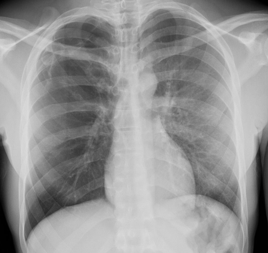
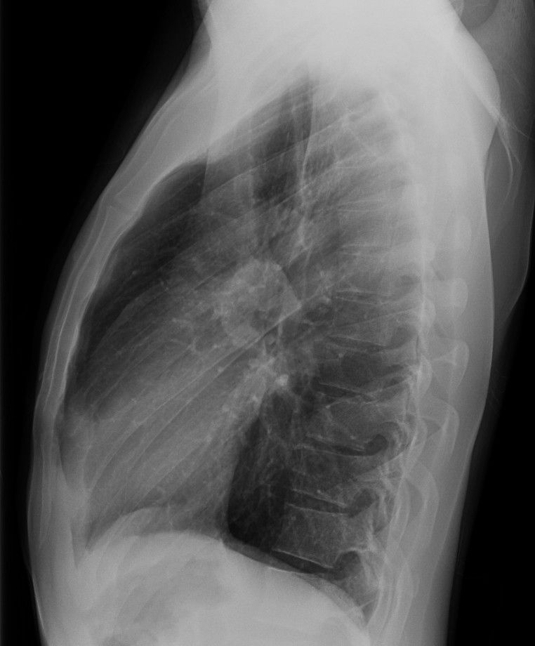
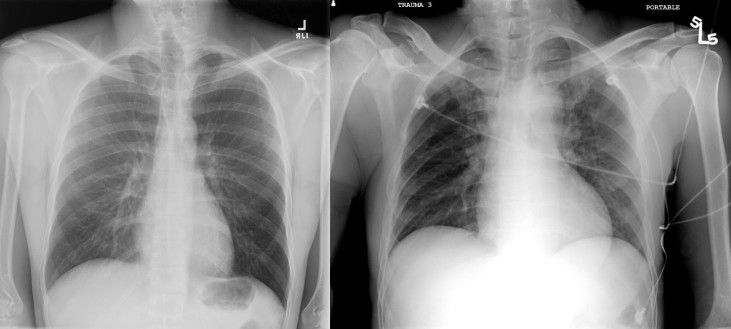
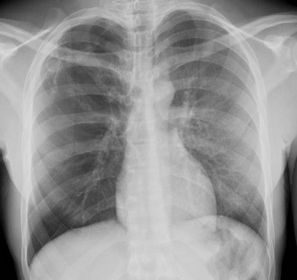
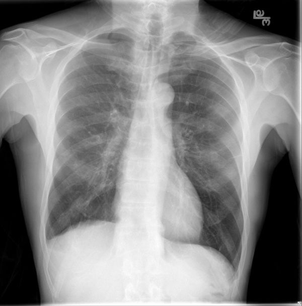
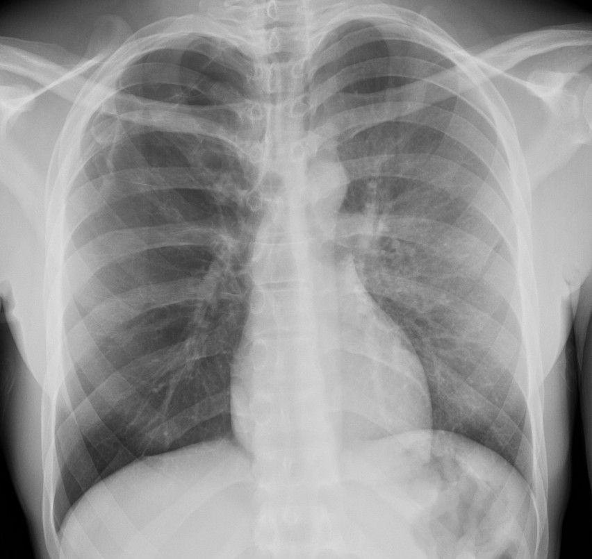
While the right hemithorax is not necessarily smaller than the left, it is definitely hyperlucent. And note the signficiantly diminished vascular markings on the right versus the left.
Oh yeah, and there is just happens to be an irregularly shaped (versus multiple) lucency in the right pulmonary apex. Bullae? Scarring? Pneumothorax?


CT from the same patient shows:
- Hyperlucent areas throughout the right lung, especially the RUL (axials - top row) and posterior RLL (coronals - bottom row)
- Bronchiectasis and bronchial wall thickening most prominent in the RUL
- Diminished vascularity throughout much of the right lung when compared to the left
- Definite pleural air (ie pneumothorax) in the right pulmonary apex with areas of scarring extending from the visceral to parietal pleura. Could this be chronic?
While our patient may not demonstrate the classic "small" lung, it does show the unilateral hyperlucent lung (CXR) and regional areas of hyperlucency/air-trapping with associated dimished vascularity and bronchiectasis consistent with Swyer-James Syndrome.
Oh yeah, and a right apical pneumothorax!
Do you want to see additional causes of Unilateral Hyperlucent lung?
Check out:
