Season 9 Case 18
History: Chest Pain

Answer:
CLICK HERE FOR ANSWER
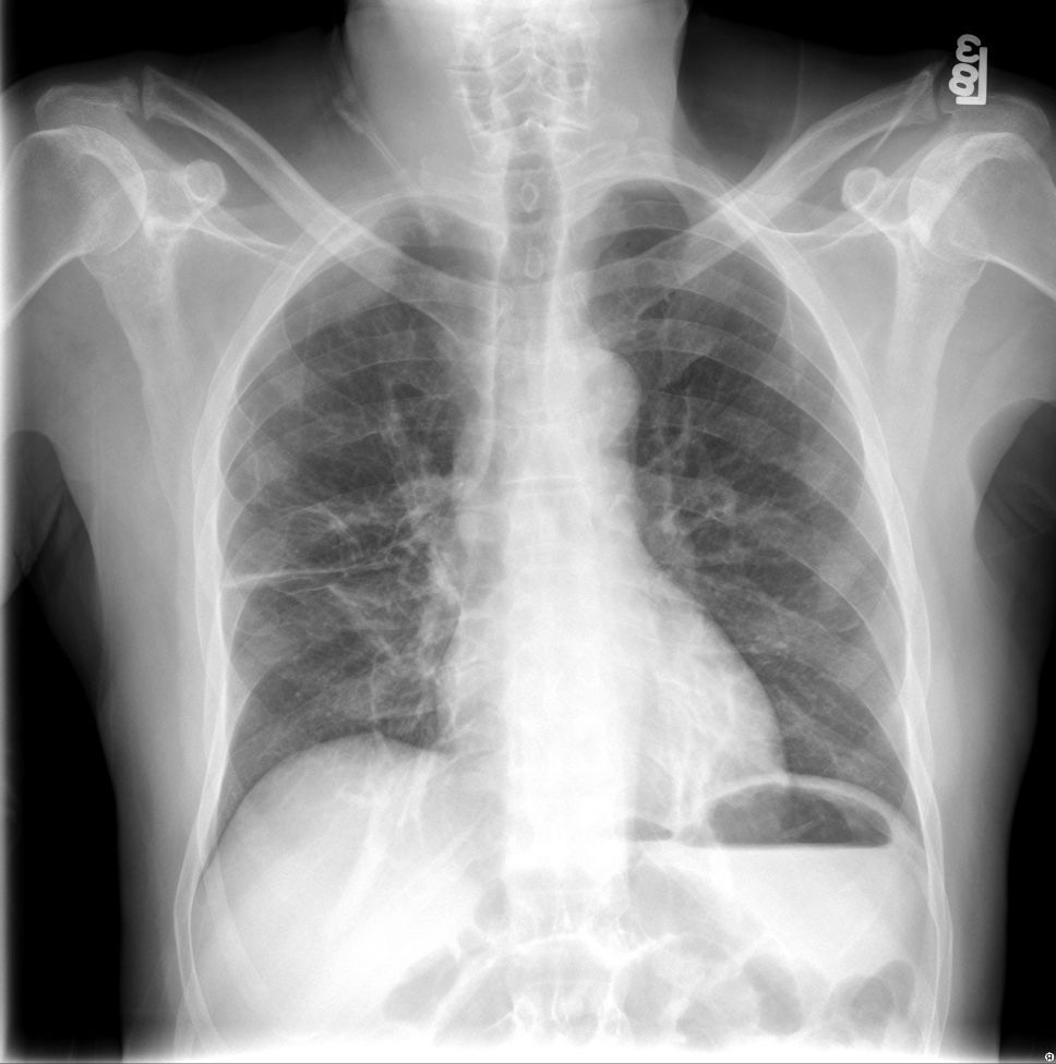
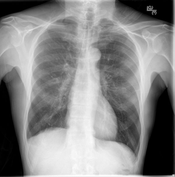
So hopefully everyone saw this pleural based opacity in the RUL. But does anyone see the other finding?

Turns out pt had some pain radiating to their neck so they got this C-spine X-ray. Perhaps one of the rare times a non-trauma C-spine X-ray is helpful?

Answer: Pancoast tumor (aka superior sulcus tumor)
RUL Pleural density is a rib met from the destructive tumor in the R pulmonary apex. Where is Right C7 TP?
These can be VERY difficult to see so you have to examine the apices closely on all CXR!


Pancoast tumor:
-varying tumor types (NSCLC m/c)
-classic hx is Pancoast Syndome (chest pain, C8-T2 radiculopathy, Horner's syndrome) but only ~25% have all 3
-often involves brachial plexus and subcl vessels to trt is radx/chemo +/- resection
-poor 5yr survival
Pancoast Tumor (Imaging)
CXR: soft tissue mass pulm apex. Look for bone destruction!
CT: improved resolution over CXR
MRI: Look for nerve/vessel involvement (if >C8 nerve involvement may be inoperable)
PET/CT: nodal, distant mets
My rec is to always examine and re-examine the lung apices, with a magnifying glass!
Look for:
- Soft tissue density (scarring v mass)
- bone destruction
- nodules
- PTX!!
***HINT: you often get a better look at the apices on the coned down C-spine CT than on the full FOV Chest CT


