Season 10 Case 18
History: Abdominal Pain
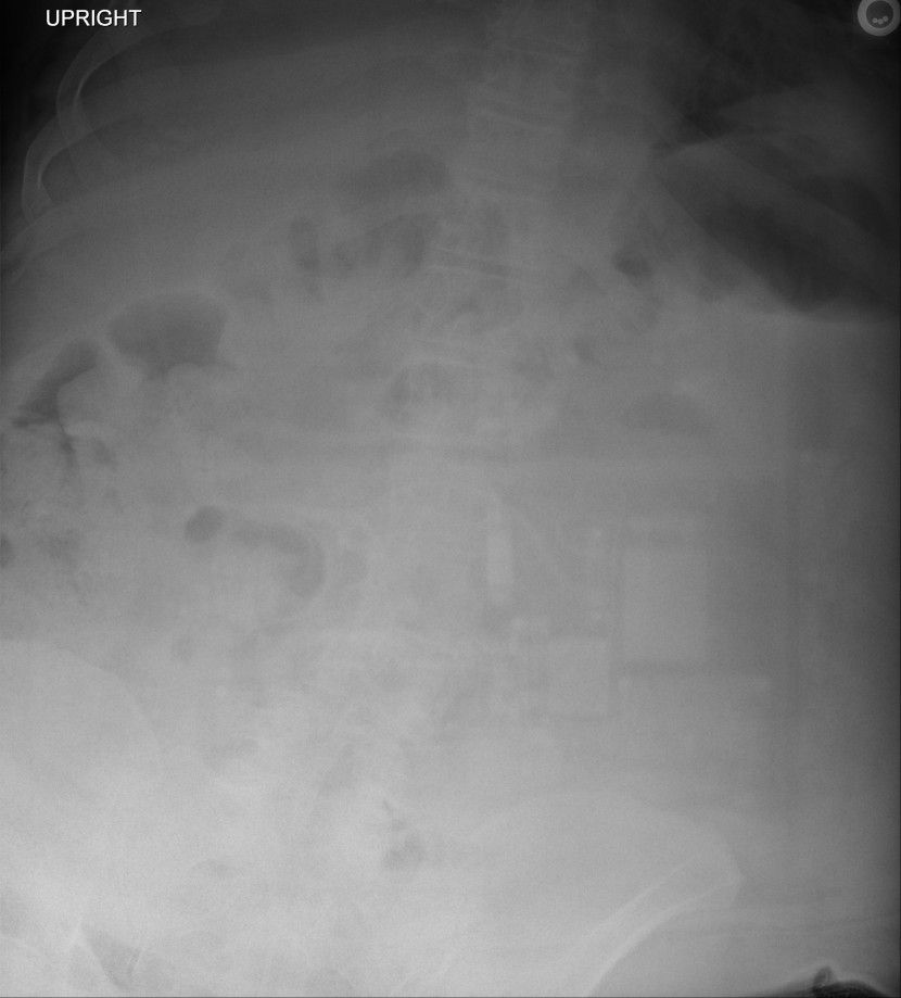
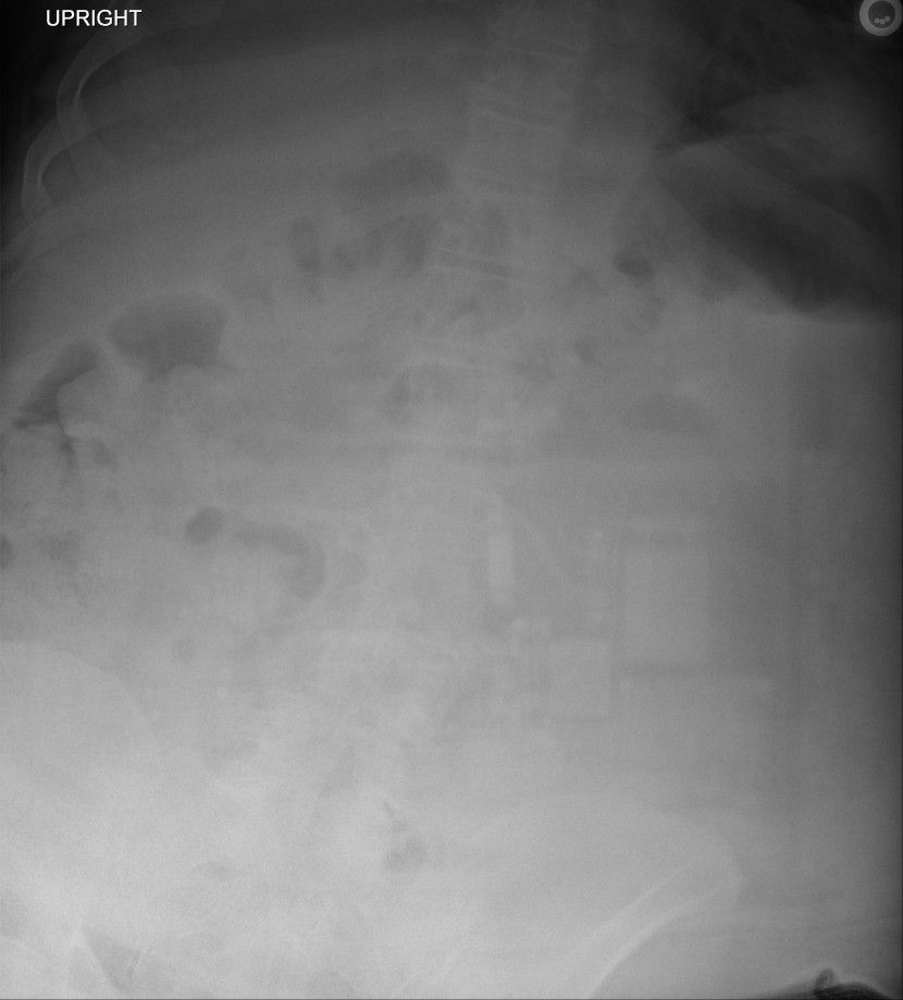
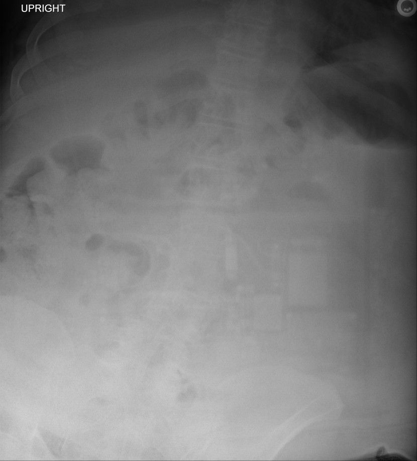
Anything wrong with this picture?
Answer:
CLICK HERE FOR ANSWER
Answer: Backscatter Artifact
There are numerous types of imaging artifact in plain film radiography. Radiopaedia has a nice compilation here:

In our case, we see metallic radiodensities overlying the abdomen that are definitely suggestive of an electronic device. But where is it? Did the patient swallow something? It is rather large to swallow isn't it?
What we are seeing is the actual electronic components of the image detector. But why are we seeing it on this exam and not all exams?

Brief Background
This is by no means complete and probably contains some questionable oversimplifaction, but I think it provides a good starting point for those starting out.... or even just not super interested < wink>< wink>
Radiographs are created by sending a "beam" of X-rays through a patient (or patient's body part) in order to "see" what is on the inside.
X-rays are similar to visible light except there are just on a different spot of the electromagnetic spectrum.
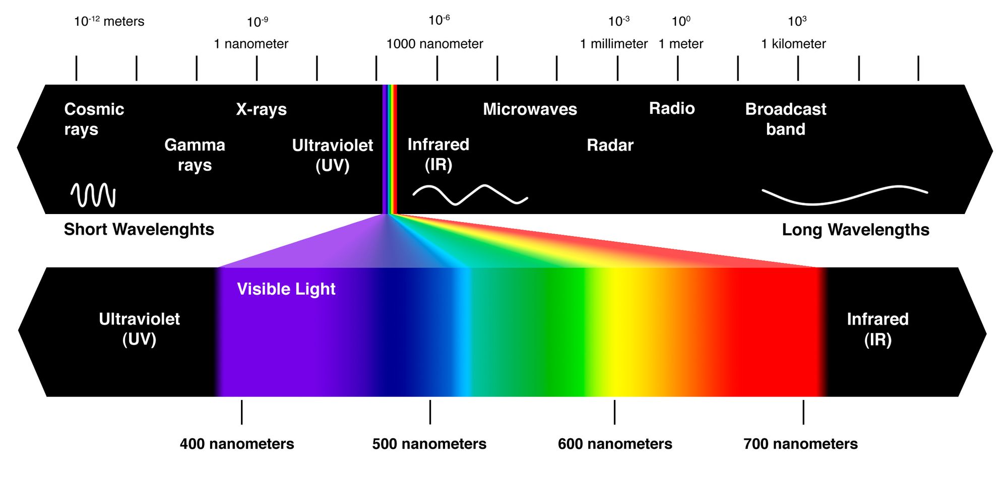
As you may know (or can see from above), the type of electromagnetic radiation changes based upon it's wavelength. And the wavelength of the radiation is inversely related to it's frequency. So as wavelength goes up, the frequency obviously goes down (there was this smart guy name Max Plank who figured this out in the early 20th century as part of we now call Quantum Mechanics).
For example:
- Broadband radio is measured in kilometers (10^3)
- Microwave radatiation is measured in micrometers (10^-6)
- Visible light is measured in 100s of nanometers (10^-9)
- X-rays are measured in single digit nanometers (10^-9)
So INVERSELY, X-rays have a HIGHER frequency, and thus a higher energy.
We can create these X-rays (often called "photons") using something similar to a light bulb. Using an electric current, we can create a stream of electrons flowing from the cathode (negative side) to the anode (positive side) of a circuit. If we stick a tungsten plate between them, the electrons will hit the tungsten and cause emission of X-rays!
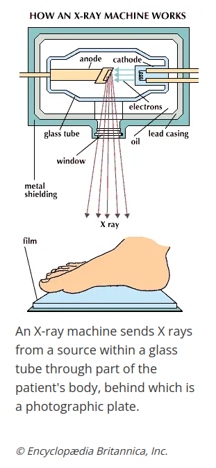
So now that we have X-rays, what do we do with them? Well shoot them at someone of course!
While visible light is relatively low energy and doesn't penetrate objects well, the higher energy X-rays can penetrate everyday material, and the degree of penetration/transmission depends upon the density of the specific material. The amount that is blocked increases (ie attenuated) as the density of the material increases (ie Metal blocks the most > bone > muscle > fat > air which blocks very little). The photons that are able to fully penetrate through the material then hit an imaging cassette that is positioned on the opposite side from the X-ray source.
Raiography Basics
The process of Radiographic imaging has evolved a great deal through the decades.
Plain-Film Radiography
Originally we had a source (X-ray generator) and a cassette that contained actual film (film-screen radiography). A short burst of X-rays are sent through the patient. The X-rays that are able to fully penetrate through the object are able to hit the film, creating silver atoms in the film emulsion which turn the film black. The amount of "blackness" is determined by the quantity of X-rays that are able to get through that one particular spot, and thus dependent on the density at that one spot.
The more dense the material, the more attenuated the X-ray beam and thus the less X-rays get through, the less silver atoms get created and the less dark the image. Air = dark and Bone = light
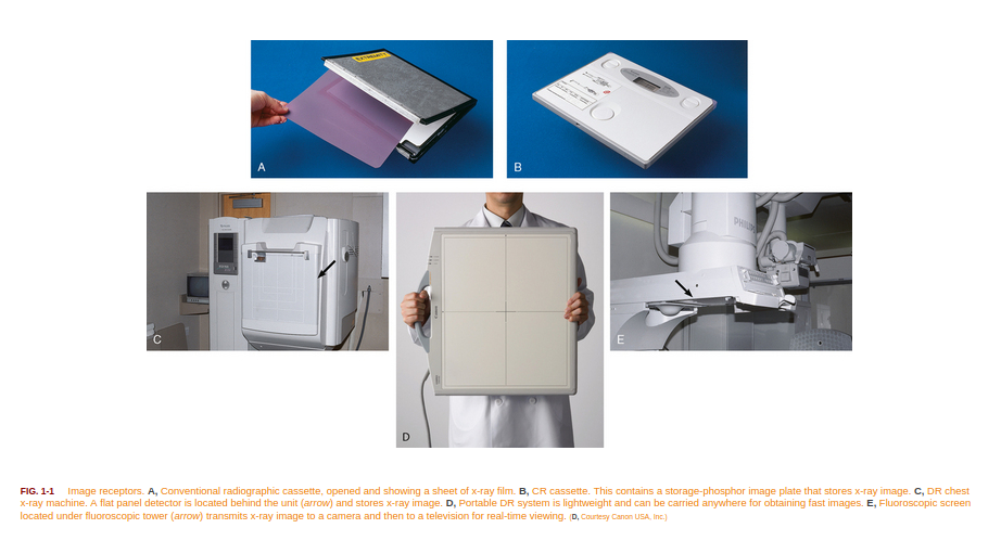
More detail can be found here. Example of the cassette can be seen in image A below.
Computer Radiography
Technological advancements lead to Computed Radiography(CR). CR replaces the film component with an image plate composed of photostimulable phosphor. When exposed to the X-ray beam, electrons are excited into a higher energy state in proportion to the amount of exposure (ie the degree of transmission and thus the material's density). We can then release these excited electrons back to normal using a technique called photostimulation where specific wavelength lasers de-excite the electrons and release their energy which can be quantified and digitized it to "digital" radiographs.
These are the the cassettes that need to be exposed and then returned to a "reader" to obtain the digital image (image B below). So while nicer than plain film (and providing better contrast and detail), we are still stuck with 1 image per cassette before we head back to the department, read and reset everything. Wanna know more?
Digital Radiography
The most recent advancement is Digital Radiography (DR). DR was able to get rid of the readers by converting the image plate to a complete flat panel detector. These dectectors can work in a couple different ways but in all forms are able to convert X-rays directly to an electric charge (ie digital signal output) (more detail here). This electric signal can be directly measured, and similar to the CR, it will be proportional to the degree of transmission and thus the density of the material.
While DR provides for even higher contrast and other imaging benefits, one of the biggest benefits is now the exposure system is entirely self-contained. Build in a wifi connection and now your "images" can be instantly sent to the PACS even if you are roaming around doing AM portable X-rays. Once sent, you can erase the "image" and reuse the same cassette over and over.(image D below)
Back to our case!
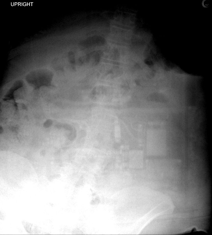
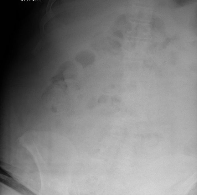
So what is backscatter artifact?
Well when the patient is larger and/or we need to cover a large area, it is necessary to increase the amount of X-rays used. Much like light that can reflect off shiny things, X-rays also refract or scatter when they hit things, going in varying directions beside just the one we want. So when these scattered X-rays actually go beyond the patient and scatter (reflect) back towards the patient, the cassette actually gets exposed from the wrong direction, with these X-rays then being attenutated (blocked) by the actual electric circuitry of the cassette itself (for easy of use, weight, etc, there is no shielding on DR cassette to block those X-rays coming from the wrong direction while there is on plain film and CR cassettes - so yes, if you see backscatter, you are using DR rather than CR or film-screen radiography).
So how can you correct for this?
- Decrease your mAs (ie drop the amount of X-rays used)
- Collimate better so more of the X-rays are focused on the detector
- Add lead backing to the cassette
Again, this is just one of the MANY type of radiographic imaging artifacts. For anyone learning or just interested, check out the Radiopaedia link above or Radiographics has a fairly good review article here:


